Muography
Proton therapy
-
Conventional proton therapy
Proton therapy is a branch of hadron therapy. It differs from conventional radiotherapy in that it uses a beam of protons rather than photons (X-rays). Radiation therapy, including both photon- and hadron-based techniques, aims to kill tumor cells. Depending on the location, size and type of tumor, different techniques are used to maximize the radiation dose delivered to the tumor while minimizing damage to the surrounding healthy tissues. Indeed, the dose that can safely be given to the tumor is limited by the need to preserve the function of nearby organs and to limit side effects for the patient.
Thanks to the physical properties of protons, proton therapy enables more conformal dose delivery, allowing higher doses to be deposited within a well-defined target volume while significantly reducing the irradiation of healthy tissues.
Figure 1 shows the dose deposited as a function of depth in tissue. Photon beams deposit their maximum dose close to the surface, followed by a gradual attenuation with depth, so that tissues located beyond the tumor are also irradiated. Protons, in contrast, release most of their energy at the end of their path, at a well-defined depth that depends on their initial energy (the Bragg peak), and deposit almost no dose beyond it. By combining several beam energies, this peak can be spread over the whole tumor volume (spread-out Bragg peak).
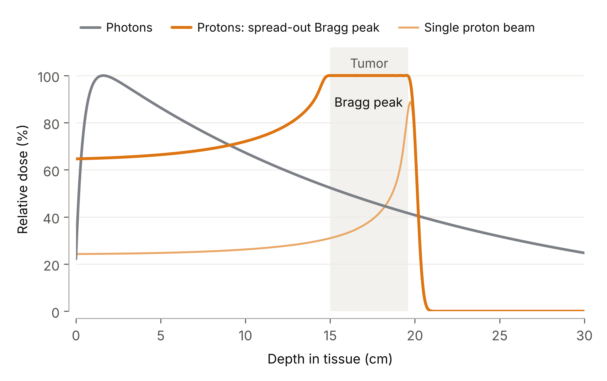
Figure 1: Relative dose as a function of depth in tissue. Photons (gray) deposit their maximum dose just below the surface and keep irradiating the tissues beyond the tumor. A single proton beam (light orange) deposits most of its energy in a narrow Bragg peak; combining several beam energies spreads this peak over the whole tumor (orange), with almost no dose beyond it. This increased precision is particularly beneficial for tumors located near critical organs or radiosensitive structures, as well as for pediatric patients. Protons are also well suited to new irradiation modalities such as FLASH therapy: unlike the low-energy electron beams used in many FLASH experiments, they can reach deep-seated tumors.
-
FLASH therapy and dosimetry challenges
In recent years, many radiotherapy and hadron therapy studies have focused on ultra-high dose rate (UHDR) irradiation delivered in short pulses. These studies have revealed an effect, now known as the FLASH effect [1]: in preclinical models, delivering the dose in a fraction of a second (~ 10 μs) spares healthy tissues while maintaining the same effect on tumors.
In conventional radiotherapy, the dose is typically delivered at dose rates of a few tens of mGy/s, whereas the FLASH effect is associated with mean dose rates exceeding 40 Gy/s, about a thousand times higher.
Clinical dosimetry, which measures the radiation dose absorbed by the patient, requires high accuracy to ensure that the delivered dose complies with the medical prescription. During treatment, dose measurements must disturb the beam as little as possible and be performed in real time, so that the beam can be interrupted immediately in the event of a problem. The most commonly used detectors that meet all these conditions are air-vented plane-parallel ionization chambers (PPICs). However, these detectors face a major challenge in the UHDR regime: the extremely high density of ions produced in the air leads to significant ion recombination and space-charge effects, so that part of the charge is never collected and the delivered dose is underestimated.
Overcoming the limitations of dose measurement under UHDR conditions, by enabling accurate, safe, and real-time dosimetry, is therefore a key challenge that will ultimately determine the clinical viability and widespread adoption of FLASH therapy.
-
Research at the IIHE
At the IIHE, we develop dosimetry methods for FLASH proton therapy within ProtherWal, the Walloon proton therapy project, in collaboration with Ion Beam Applications (IBA). To understand how ionization chambers behave at ultra-high dose rates, we extended the Garfield++ simulation toolkit, developed at CERN, with a three-dimensional Monte Carlo model of charge transport, electron attachment, ion recombination and space-charge effects [2]. These developments are now part of the official Garfield++ code (see the ionization chamber tutorial ), and the complete simulation framework, GarFLIC , is openly available.
The simulations show that space charge can locally increase the electric field in the chamber by more than a factor of four, or reduce it to almost zero. They also indicate that the loss of collected charge is mainly driven by a decrease in the fraction of free electrons, i.e. electrons that reach the electrode before attaching to oxygen molecules and therefore escape recombination. Building on these results, our next steps are to validate a correction model with beam measurements and to implement it on FPGA electronics, in order to estimate the delivered dose in real time (< 1 ms), between two successive beam pulses.
-
References
- Favaudon, Vincent, Laura Caplier, Virginie Monceau, et al. “Ultrahigh Dose-Rate FLASH Irradiation Increases the Differential Response between Normal and Tumor Tissue in Mice.” Science Translational Medicine 6, no. 245 (2014): 245ra93. https://doi.org/10.1126/scitranslmed.3008973 .
- Gérard Ortega, Pierre, Gilles De Lentdecker, Quentin Flandroy, and Jarrick Nys. “Charge Collection Efficiency in Air-Vented Plane-Parallel Ionisation Chambers at Ultra-High Dose Rates: A Self-Consistent Garfield++ Monte Carlo Model Including Space-Charge Effects and Ion Recombination.” arXiv:2606.24384 [physics.med-ph] (2026). https://arxiv.org/abs/2606.24384 .